I’ve been away in Burma with the BBC for some time looking for animals and since getting back I’ve been thinking it was high time to put together a proper website that could incorporate my blog. So, here it is, albeit a work in progress. All new posts will be on this website.
Posted by: scrubmuncher | May 7, 2013
It’s been a while…
Posted in Uncategorized
Posted by: scrubmuncher | October 26, 2012
Reproduction at its most rampant…
Parasitic platyhelminthes, particularly the flukes, have the most complex life cycles of any animal, involving two or more hosts and an elaborate sequence of developmental stages that allows each egg laid by the adult to spawn huge numbers of infective juveniles via asexual reproduction. The photo and video below illustrate a part of this developmental sequence in a fluke that uses periwinkle’s as its intermediate host.
The photo shows part of the periwinkle’s gut dissected to reveal an infestation of pale, ovoid structures. These are known as sporocysts and they’re much less than 1 mm long. Every one of these sporocysts developed from a single miracidium, a ciliated organism that hatched from an adult fluke’s egg. The video reveals the macabre secret contained within these sporocysts.

Part of an unfortunate periwinkle’s gut. This individual was infested with sporocysts (pale, ovoid structures), one stage in the life cycle of a fluke (Ross Piper).
The cercariae released from the bulging sporocysts are another infective stage in the parasite’s life cycle and it is their task to penetrate the body of the definitive host (unknown in this case) where they develop into the adult fluke. For a fluke, the example above is a relatively simple life cycle. In other species there are yet more body forms and serial repetition of some stages to produce colossal numbers of cercariae from a single egg. What is the significance of this singular reproductive strategy? Well, in short, huge numbers of offspring produced asexually offset the huge odds stacked against any one juvenile completing its development. You have to love the incredible complexities of such animals that are often disregarded as ‘simple’.
Posted in Bewildering diversity | Tags: asexual reproduction, fluke, life cycle, parasite, platyhelminthes, trematode
Posted by: scrubmuncher | October 16, 2012
Magnificent mouthparts
The Stenus rove beetles have featured in a couple of other posts, but the photos below are worthy of revisiting these fascinating little insects. I had these images done by a chap who has a SEM in his converted garage. These are only small versions of the original images, bu if you click on them you’ll see the detail is incredible, especially the last one that shows the tip of the beetle’s labium with numerous sensory structures. These adhesive surfaces stick fast to the prey, yanking it back to the sickle-like mandibles of the beetle as the labium is withdrawn.

Stenus clavicornis with its telescopic labium (poking out from between the mandibles) revealed for the camera (Ross Piper and David Spears).

Stenus clavicornis – close up of the head and labium. Note the cruel mandibles too (Ross Piper and David Spears).

Stenus clavicornis – close-up of the tip of the labium. Just look at how elaborate this tiny structure is (Ross Piper and David Spears).
Posted in Admirable adaptations | Tags: carnivorous beetle, feeding adaptations, insect mouthparts, mouthparts, rove beetle, Stenus
Posted by: scrubmuncher | October 12, 2012
Time travellers
When I was working on the book I’ve just finished I became a little fixated by the rotifers and I blogged about them previously, here and here. However, these two posts are not enough and I feel like I must discuss these tiny creatures at length such is their mind-boggling biology. One thing that particularly fascinates me about the rotifers and the other small animals of transient aquatic habitats is their ability to time travel. I don’t mean they have a glowing, whirring device at their disposal that transports them to a year of their choice, but they do have adaptations that are perhaps the next best thing. Namely, by entering a state of very deep quiescence (cryptobiosis) or persisting as resting eggs they slow their vital processes to such a degree that the spark of life is vanishingly faint – perhaps 0.01% of normal. This allows them to cross stretches of time effectively unaged between periods of favourable conditions when they can get on with feeding and reproducing. Exactly how long they can survive these periods of suspended animation is a bone of contention, but it’s certainly decades and possibly even centuries or millenia. Below are some photos illustrating these adaptations in rotifers and tardigrades.

The rotifer, Pompholyx sulcata. Attached to the rear-end of this rotifer is a large resting egg – a time-travelling adaptation of many rotifers species (Image courtesy of Michael Plewka).

The resting egg of a tardigrade. These eggs have a thick shell that is very often elaborately sculpted (image courtesy of Eye of Science/SPL).

A tardigrade entering the state of cryptobiosis. This complex dehydration process involves all the water molecules in the body of the animal being replaced with a simple sugar, typically trehalose (image courtesy of Microfield Scientific Ltd /SPL).

Following on from the image above, the process of cryptobiosis is almost complete and the tardigrade is now little more than a dried out husk (a tun) that can see out very long periods of unfavourable environmental conditions (Image courtesy of Eye of Science / SPL).
Posted by: scrubmuncher | October 12, 2012
Unfeasibly large gonads…
This post isn’t about the famous Viz character, rather the mind-boggling diversity of nematodes, which I touched on in a previous post. One nematode that beautifully illustrates the bizarre life histories of these animals is Sphaerularia bombi, a parasite of queen bumblebees. Like so many other parasites this little nematode manipulates the behaviour of its host in order to increase its chances of continuing the freeloading tradition. In short, a queen bee afflicted with this nematode forages and feeds, but she makes no effort to found a nest of her own. Instead, she is compelled by the parasite to return to the place where she hibernated or a another suitable spot and it is here the nematode’s offspring find their way into the outside world in the faeces of their host where they wait to infect another queen bee that makes use of the hibernaculum.
Such a strategy is a long shot to say the least, so to offset the long odds huge numbers of offspring are required. With a body only a few millimetres long the nematode is rather limited in how many eggs it can produce, but to get around this problem something incredible happens to its gonads. Its entire reproductive system turns inside out and balloons to relatively enormous proportions, eventually dwarfing the nematode that dangles at the end of this structure like a vestigial appendage (see photo below). With unfeasibly large gonads the nematode can produce tens of thousands of eggs.

Dwarfed by its enormous, inside out uterus, the nematode, Sphaerularia bombi, can be seen near the centre of this image (image from Mark Blaxter’s teaching pages).
Posted in Admirable adaptations | Tags: bee parasite, bumblebee, nematode, nematode diversity, parasite, parasitic nematode, sphaerularia bombi
Posted by: scrubmuncher | September 23, 2012
Pin head
Last year in northern Spain we crashed our hire car, but after exercising our rudimentary knowledge of the Spanish language we eventually found a friendly mechanic to repair it. The next day we had to walk the 17 kms from Pesaguero to Potes along an old track through the mountains to pick up the repaired motor. It turned out this long and very hot yomp was well worth it as a splendid variety of beasts were espied, including this fine, little wasp.
This is another type of predatory, solitary wasp, but this species exhibits an incredible degree of sexual dimorphism. The individual in the photo below is a male and compared to the female (see here for the female of a related species) he’s a veritable pin-head. Apart from his odd-shaped head the male also has very distinctive, enlarged fore-legs, perhaps for signalling to the female and gripping hold of her when his courtship has been well-received. Active hunters, female Lestica wasps stock their nests with small moths. The dandy males, on the other hand, simply spend their time looking for females and sipping nectar.

A pin-headed male Lestica sp. Sexual dimorphism is very distinct in this genus of solitary, predatory wasps (Ross Piper).

Male Lestica spp. have a very odd-shaped head and large, flattened fore-limbs (Ross Piper).
Posted in Bewildering diversity
Posted by: scrubmuncher | September 19, 2012
Mini-hawks
In one of my older posts I banged on about the hunting prowess of some of the solitary wasps and I alluded to the visual acuity of these mini-hawks.
This post concerns Ectemnius wasps in particular. These have to be right up there amongst my favourite animals. If you’ve ever observed these wasps you can’t help feeling they’re rather smart. Sharp-eyed, they watch their surroundings intently, their little heads flicking this way and that in search of mates, prey and enemies.
To give you a better idea of just how good the vision of these insects is I took the liberty of photographing their heads through a microscope. You’ll see from the photos below that their eyes are relatively enormous and are composed of hundreds of individual facets (ommatidia). There’s no question that eyes of this type are second to none when it comes to detecting movement, but they must also be able to form pretty decent images if the fussy dietary requirements of these wasps are anything to go by. Ectemnius wasps prey on flies, typically hoverflies, but also muscids, etc. which are masters of the air in their own right, so hunting them, often on the wing, requires an amazing coordination of flight and vision. The images formed in the brains of these wasps from the information streaming in through their compound eyes must be sufficiently detailed to allow them to single out their prey from inanimate objects and other similarly sized insects.

The massive compound eyes of a predatory solitary wasp (female Ectemnius cephalotes). Note the fine silvery ‘moustache’ (Ross Piper).

If you were a hoverfly you would not want one of these bearing down on you. The fixed gaze of a female Ectemnius continuus (Ross Piper).
Posted in Admirable adaptations | Tags: compound eyes, ectemnius, insect vision, predatory wasps, solitary hymenoptera, solitary wasps
Posted by: scrubmuncher | September 7, 2012
A grisly end for an earthworm…
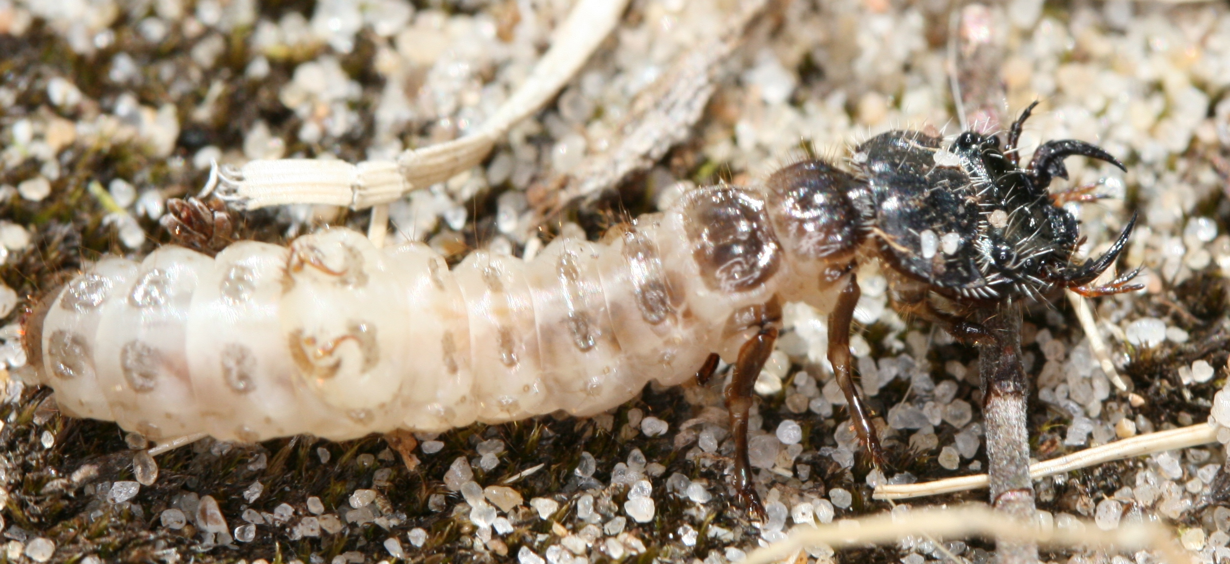
Whilst camping in Cornwall recently we found this earthworm thrashing about in the turf. I thought nothing of it initially, but on closer inspection the poor worm was writhing about to shake off an attacker, none other than the larva of a flesh fly that had sunk it’s mouth-hook into the body of the unfortunate annelid and was rending its flesh. The earthworm squirmed and coiled, but the fly larva was in no mood to relinquish its grip and before long the struggle continued back down in to the grass with the fly larva as the probable victor.
This chance observation really goes to show how many struggles and interactions go on unseen under our noses. Typically, sarcophagid (flesh flies) larva eat decomposing animal matter, but perhaps many of them have more specific tastes, actively hunting out other animals to feed on?

A flesh fly larva predating an earthworm (Ross Piper).

Try as it might the earthworm just couldn’t shake this little predator (Ross Piper).
Posted by: scrubmuncher | September 5, 2012
A tiger beetle on steroids
Tiger beetles come in all shapes and sizes, but on the whole they’re rather elegant, fine-limbed animals. With this said, some of their number are real brutes and perhaps the most brutish of all are the African Manticora species (for a sense of scale here’s one behaving nicely on someone’s hand).
Massive and completely black, they scuttle around in the dead of night chomping any suitably sized animals that venture within range of their enormous mandibles. Unlike most their kind they’re completely flightless and relatively cumbersome. The fine specimen in the photo below was found at a campsite we stayed at in Namibia at the end of 2011. It was lurking under the low vegetation growing around a lamp-post, idly waiting for other insects drawn by the light to lose their bearings and fall to the ground.
Getting close enough to most tiger beetles to take decent photos is a bit like herding cats because they’re just so fast and skittish. However, these beefcakes of the tiger beetle world are much more relaxed and this one happily made a small snack of several flying termites while I had my camera in its personal space. After growing weary of the flash the beetle scuttled off like a wind-up toy. The next challenge is to find the fully-grown Manticora larva. It would look a bit like this, only much much bigger.
If you want to find out more about these splendid insects then this paper is packed with information. There are even some photos of the formidable larvae, their tunnels and their parasitoids.

The African Manticora spp. are the biggest and meanest looking of all the tiger beetles. Thirteen Manticora spp. are currently known from throughout sub-Saharan Africa (Ross Piper).

Here’s the same specimen unceremoniously dispatching a winged termite (Ross Piper).
Posted by: scrubmuncher | August 15, 2012
Microfauna
[This post is adapted from a book I’ve just finished, which will be published sometime next year by Thames and Hudson.]
Animals come in a huge range of shapes and sizes, but the vast majority of them are very small. In fact there are many animals that are way smaller than some single-celled organisms.
Here, on the right is a rotifer, an animal with a body made up of around 1,000 cells. At the top, contracting rapidly, is a ciliate, a single-celled organism (Spirostomum spp.). The tip of a needle gives a sense of scale. Disappearing behind the needle at the start of the video is another single-celled organism.
It is creatures like this that really underline the multi-layered complexity of biodiversity. These cilates, swimming around in a small pond, are little bigger than a full stop yet they dwarf the rotifers that crawl and dart around them. Humans on the other hand are big organisms, vastly bigger than most other animal species, but this smaller majority is effectively hidden in plain view, which makes it difficult to appreciate the riot of animal life that surrounds us.
Posted in Uncategorized | Tags: ciliate, microscopic animals, pond life, rotifer, rotifers, single-celled organism, spirostomum
